The Lyceum: Brain & Mind Weekly — Mar 21, 2026
Week of March 21, 2026
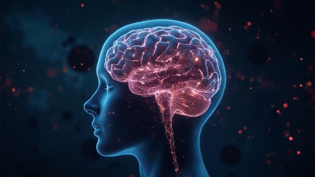
The Big Picture
This was a week of uncomfortable revelations about what we thought we understood. TMS — the workhorse brain stimulation therapy for depression — turns out to be activating neurons nobody expected, in patterns nobody predicted. Astrocytes are co-authoring your memories. A hidden peptide loop is reshaping the reward circuit from the inside. And the mind-reading conversation lurched forward: implanted electrodes can now decode what you're about to decide before you know you've decided it. Individually, each finding is significant. Together, they paint a picture of a brain that is far less modular, far less neuron-centric, and far less private than the textbooks suggest.
This Week's Stories
You've Been Stimulating the Wrong Neurons — and Now We Can See Which Ones Actually Fire

Transcranial magnetic stimulation has been FDA-cleared for depression for years. The pitch: pulse a magnetic coil at the prefrontal cortex — the brain's executive region behind your forehead — and somehow, weeks later, mood improves. The "somehow" has always been the embarrassing part. Nobody knew what was happening at the single-neuron level in the deep structures that actually regulate mood.
A preprint posted March 18 changed that. Using microelectrode recordings in four neurosurgical patients, researchers captured single-neuron spikes as early as 8 milliseconds after a TMS pulse to the left dorsolateral prefrontal cortex — a window nobody has seen through before. TMS triggered time-locked responses in 46% of recorded neurons in the study across deep structures including the striatum and thalamus on both sides of the brain. The key surprise: TMS preferentially activated inhibitory interneurons (local circuit brakes) in these regions while suppressing excitatory pyramidal cells on a slower timescale.
What this means: TMS may work for depression not by boosting the prefrontal cortex, as usually assumed, but by quieting an overactive emotional circuit from above. If this holds, it opens the door to designing stimulation protocols that target specific cell types rather than a skull location — potentially making TMS faster and more reliable. If it doesn't replicate, we're back to guessing. The signal to watch: whether follow-up studies in larger cohorts show the same inhibitory-first, excitatory-second pattern, and whether anyone designs a clinical protocol around it.
A related methodological finding deserves attention: a separate multisite TMS-fMRI study found that stimulation-induced discomfort accounts for 12% of TMS-evoked brain responses in that multisite study — and 25% in those with depression or anxiety in that study. In the populations most likely to receive TMS therapy, up to a quarter of what researchers measured as "treatment signal" may have been pain signal all along.
Synapses Caught Mid-Fire at Near-Atomic Resolution

What happens the instant a neuron sends a signal? We just got the sharpest picture yet. Using cryo-electron tomography — a technique that flash-freezes brain tissue and images it in 3D at near-atomic resolution — researchers mapped the exact protein arrangement in synapses poised for neurotransmitter release. Published in Nature, the images reveal a scaffolding structure around neurotransmitter vesicles (the tiny packets of chemical signals) that constrains release timing more tightly than existing models assumed.
What changes: Every computational model of how synapses strengthen or weaken — the molecular basis of learning — now has new structural constraints to incorporate. If modelers adopt these parameters, expect a wave of re-calibrated plasticity simulations this year. If the field treats this as a pretty picture and moves on, it's a missed opportunity. Watch for whether computational neuroscience groups cite this in updated synapse models by summer.
The Reward Circuit Has a Hidden Peptide Loop That Reshapes Everything Around It
The ventral tegmental area — a cluster of dopamine neurons deep in the brainstem — is the engine of your brain's reward system. The standard story: these neurons release dopamine to signal salience and reinforce behavior. But dopamine neurons are also being tuned from within, by signals most researchers haven't tracked.
A preprint posted March 16 reveals that the neuropeptide cholecystokinin (CCK) is released not from nerve terminals but from the cell body and dendrites — the receiving end — of dopamine neurons themselves. When a single dopamine neuron fires, CCK simultaneously strengthens inhibitory connections and weakens excitatory ones on that same neuron. More strikingly, this effect spreads: activating one dopamine neuron reshapes synapses on neighboring neurons up to 100 micrometers away.
What changes: This means a single neuron can rewrite the synaptic landscape across an entire local dopamine population at once — a previously unappreciated layer of reward circuit control. If this mechanism operates during real behavior (this is mouse brain-slice work, not behaving animals), it could explain how reward signals get amplified or dampened in addiction, motivation, and stress. The failure mode: if CCK release doesn't scale the same way in intact, behaving circuits, this stays an elegant slice finding. Watch for optogenetic experiments testing CCK's role during actual reward-seeking behavior.
Implanted Electrodes Can Now Decode Your Decisions Before You Make Them

Brain-computer interfaces have always been framed as communication tools — helping paralyzed patients type or speak. This week, a Nature analysis piece reframed the conversation in more unsettling terms.
An implanted-electrode study reported preconscious decision decoding up to approximately two seconds before subjects reported awareness of their choice, using machine learning on intracranial recordings. Separately, a non-invasive EEG study showed that even scalp-level recordings combined with machine learning could predict which category of image a person would perceive milliseconds before they reported seeing it, with around 80% accuracy in that cohort.
What changes: The gap between "decoding what someone said" and "reading what someone is about to decide" has collapsed faster than anyone expected. If these results replicate at scale, they force a legal question that doesn't have an answer yet: does preconscious thought deserve legal protection? The EU AI Act is already being extended to neurotechnology, and several U.S. states are considering cognitive privacy legislation. If regulators move slowly, commercial applications may outpace the law. The signal: watch whether any federal agency issues guidance explicitly distinguishing invasive from non-invasive mind-reading within the next six months.
China Quietly Approves the First Commercial Brain Implant for Paralysis
While U.S. headlines focus on Neuralink demos, China did something more concrete: it reportedly approved the first commercial brain-computer interface implant to restore hand movement in people with paralysis. The device — a semi-invasive system placing electrode arrays on or just above the sensorimotor cortex — sends signals wirelessly to external decoders. In prior clinical work at Tsinghua University, patients with long-standing tetraplegia achieved over 90% grasp accuracy in those studies after several months of training.
South China Morning Post described an approval for Neuracle Medical Technology — a coin-sized wireless implant driving a robotic glove. Reddit threads flagged a separate approval under the name Borui Kang Medical. Whether these represent one device or two, the regulatory signal is the same: a country-level health system has decided BCIs are ready to be reimbursable medical technology for paralysis, not research prototypes.
What changes: That development comes amid pressure for U.S. and EU regulators to clarify their own commercial pathways. If China publishes post-market surveillance and safety data that hold up, it accelerates global adoption. If adverse events emerge without transparent reporting, it sets the field back. Watch for Chinese regulatory labeling requirements and whether Western agencies respond with timeline commitments.
Your Astrocytes Are Co-Writing Your Memories
The neuron-centric model of memory — where neurons are the authors and everything else is scaffolding — took another serious hit this week. A Perspective in Nature Reviews Neuroscience consolidates evidence that astrocytes, the star-shaped glial cells long dismissed as support structures, participate in memory traces through coordinated activation in sparse ensembles. Researchers are calling these "astro-neuronal engrams" — the physical trace of a memory co-authored by neurons and the glial cells surrounding them.
Complementing this synthesis, rodent experiments using optogenetic manipulation of hippocampal astrocytes showed that astrocyte calcium waves can promote synaptic strengthening and accelerate maze learning — a more causal demonstration that astrocytes actively shape plasticity.
What changes: Every memory-enhancing drug, every Alzheimer's therapy, every model of age-related cognitive decline is built on the assumption that neurons are the target. If astrocytes are co-writers, therapies ignoring them work with half the script. The failure scenario: if astrocyte contributions turn out to be modulatory rather than essential, the neuron-first approach survives. The signal: watch for whether Alzheimer's drug trials begin including astrocyte biomarkers as secondary endpoints.
Synthetic Cells Form Memories — Without a Single Neuron
Michael Levin's lab at Tufts published a preprint showing that Xenobots — tiny, self-propelled entities made from frog embryo cells with no nervous system and no genome editing — form long-term, stimulus-specific memories. After brief exposure to different chemical stimuli, the Xenobots showed distinct behavioral changes detectable through both gene expression and physiological signatures.
What changes: If confirmed, this challenges the assumption that memory requires neurons. Information storage may be a property of cellular machinery that nervous systems co-opted, not invented. The failure mode is familiar for Levin's lab: ambitious interpretations of Xenobot behavior have drawn lively debate before, and the cellular mechanism — how chemical exposure produces lasting transcriptional changes — is still being worked out. This is a preprint, not peer-reviewed. But the question it raises is real and testable: what is the minimal biological substrate for a memory?
NIH Bets $15 Million That Brain Organoids Can Replace Animal Testing
Brain organoids — small 3D clusters of neurons grown from human stem cells — have been proof-of-concept curiosities for years. A new NIH grant signals the field is serious about making them infrastructure.
Johns Hopkins received a five-year, $15 million grant to build DROIDp (Drug Research Organoid Intelligence Development Platform), which will use brain organoids, advanced electrical sensors, and AI analytics to assess neural functions like learning and memory for drug and chemical testing. The platform will evaluate organoids from healthy individuals and patients with Alzheimer's and SYNGAP1-related disorders — a rare pediatric condition involving intellectual disability and seizures. The FDA has previously indicated it would promote organoid use as an alternative to animal testing.
What changes: Organoids are moving from research tool to regulatory-adjacent infrastructure. If DROIDp demonstrates that organoid "learning" predicts human drug response, it could reshape how neurological drugs are screened — faster, cheaper, and without animal behavioral tests. If organoid readouts don't correlate with human outcomes, the platform becomes an expensive curiosity. Watch for validation studies comparing organoid predictions to clinical trial results within two years.
Building Muscle May Be a Targeted Antidepressant — Especially for Women
Exercise and depression is well-trodden territory, but new evidence this week adds specificity the usual headlines miss. A Mendelian randomization study — which uses genetic variants as natural randomized experiments — analyzed up to 341,326 UK Biobank participants and found that genetically predicted higher muscle strength was associated with lower odds of depression. Genetically predicted cardiorespiratory fitness did not show the same effect. The association was stronger in women than men.
The mechanism isn't endorphins. Muscle tissue secretes myokines — signaling proteins including irisin and IL-6 in anti-inflammatory mode — that cross the blood-brain barrier and interact with BDNF (brain-derived neurotrophic factor, a protein supporting neuron survival and plasticity). Strength training also appears to dampen HPA axis dysregulation — the stress-hormone system consistently implicated in depression. Complementary reporting confirmed the genetic causal inference approach strengthens the case beyond correlation.
What changes: If replicated in non-European and younger cohorts, expect depression guidelines to start naming progressive resistance training alongside psychotherapy and SSRIs — with sex-specific recommendations. If the sex difference doesn't hold, the prescription stays generic. Watch for intervention trials that manipulate strength precisely, not just encourage general exercise.
New Products & Launches
- Cirrus AI Brain-Mapping Tool received FDA market authorization for pre-surgical neural planning. If hospitals adopt it for routine neurosurgery prep, it could quietly compress clinical neuroscience workflows. Early adoption signals from surgical centers will indicate whether AI-assisted brain mapping becomes standard or stays niche.
- DROIDp Organoid Platform (Johns Hopkins / NIH): While not commercially available yet, the $15M grant formally launches a drug-testing infrastructure built on brain organoids and AI readouts — the first federally funded platform explicitly designed to replace animal behavioral testing for neurological drugs.
⚡ What Most People Missed
- The "social brain" may not be social at all. A Nature Reviews Neuroscience Perspective argues the medial prefrontal cortex handles social cognition not because it's a dedicated social module, but because social prediction is one instance of a broader inference computation. If this reframing sticks, it reorganizes how we model — and treat — social deficits in autism and schizophrenia.
- TikTok is functioning as an unregulated ADHD textbook. Multiple independent analyses converge: roughly 52% of ADHD-related TikTok videos in those analyses contain inaccuracies, and exposure to this content reduces factual knowledge while increasing confidence in that knowledge — the most dangerous pattern in health misinformation. Public health responses to neurodevelopmental disorders will increasingly need to be platform-aware.
- The COSYNE 2026 funding alarm. When the chairs of the COSYNE conference open by discussing the collapse of basic science infrastructure — shrinking labs, evaporating postdoc positions, unprecedented federal funding pressure — that's a signal about the pipeline from discovery to clinical translation, not just academic politics.
- DNA folds like origami to control which neuron you become. Daniele Canzio's lab at UCSF discovered that chromatin folding patterns act as molecular barcodes maintaining neuronal identity across decades. Failures in these patterns may underlie autism, schizophrenia, and Alzheimer's — a mechanism that's now testable but hasn't hit mainstream coverage.
- Non-hallucinogenic neuroplastogens are entering preclinical pipelines. Tabernanthalog (TBG), an ibogaine analog, promotes neural rewiring without hallucinations or cardiotoxicity. If this drug class translates to humans, it changes the access model for circuit-rewiring therapies from supervised psychedelic sessions to take-home pills.
📅 What to Watch
- If Chinese regulators publish post-market surveillance requirements for the newly approved BCI implants, it will define what "acceptable risk" looks like for movement-restoring neural devices worldwide — and force Western agencies to respond with their own timelines.
- If the single-neuron TMS findings replicate in larger cohorts and someone designs a clinical stimulation protocol targeting the inhibitory-first pattern, it could make depression treatment with TMS both faster and more predictable — the first mechanistically informed protocol redesign in the therapy's history.
- If the AD/PD 2026 conference preprints confirm that lecanemab's Fc fragment is the functional heart of amyloid immunotherapy, every Alzheimer's antibody in development will need to be re-evaluated against that architecture — reshaping a multi-billion-dollar pipeline.
- If large platforms respond to the converging TikTok ADHD misinformation coverage with actual ranking changes (boosting clinician-vetted content), we get a live experiment in whether algorithm tweaks can measurably reduce misinformation exposure and alter self-diagnosis or help-seeking patterns at population scale.
- If organoid "learning and memory" readouts from DROIDp correlate with human drug responses in early validation studies, it marks the moment brain organoids cross from curiosity to regulatory infrastructure — with implications for every neurological drug in development.
The Closer
A frog cell remembering a chemical it met once, a magnetic pulse accidentally quieting the wrong neurons for the right reasons, and a country approving a brain implant while the rest of the world is still arguing about the paperwork.
Turns out the biggest confound in depression treatment was that the therapy hurt — and nobody thought to subtract the ouch from the data.
Until next week, keep your astrocytes in the conversation.
If someone you know would enjoy learning that their glial cells have been ghostwriting their memories, forward this to them.
From the Lyceum
The FTC just moved AI hiring bias from ethics panels to active federal enforcement — a shift that matters for anyone thinking about how algorithms shape human outcomes at scale. Read → The FTC Just Made AI Hiring Bias a Federal Enforcement Target